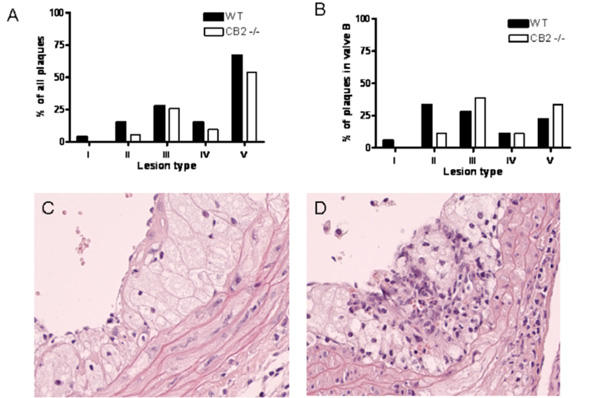
Fig. (4) A) Lesion severity in the aortic valve area presented as percentage of all plaques (I. early fatty streak, II. regular fatty streak, III. mild plaque, IV. moderate plaque, V. severe plaque). B) Lesion severity of the plaques in valve B as percentage of all plaques in valve B. Representative photomicrographs of an atherosclerotic lesion containing: C) large foam cells or D) inflammatory infiltrates with small macrophages stained with haematoxylin-eosin (magnification 40x).