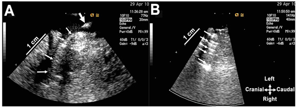
Fig. (2A) A balloon catheter with a coil on its tip has been placed into the right renal artery (small arrows) via the introducer catheter whose
tip was subsequently pulled back (thick arrow). (B) The balloon catheter was inflated to deploy the coil, and after deflation pulled back from
the renal artery lumen. The ultrasound scan shows reflections (arrows) from the coil.