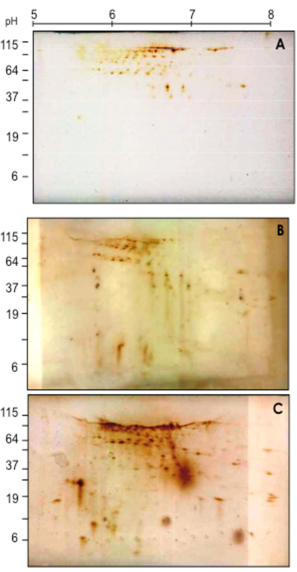
Fig. (2) 2D-SDS Western blotting patterns of biotinylated coronary VELM proteins during control (A), Ischemia (B) and I/R (C). VELM proteins were biotin labeled during control conditions. Notice that from A to B to C there is an increase in the number of low molecular weight spots.