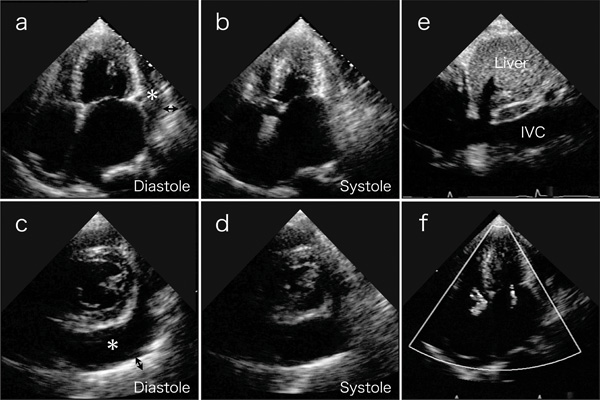
Fig. (2) Transthoracic echocardiography on 4-chamber views (a and b) and short axis views (c and d) reveal a large amount of circumferential pericardial effusion (asterisks), thick visceral pericardium (two-headed arrows), left ventricular apical myocardial hypertrophy with normal contraction, and bilateral severe atrial dilatation. (e) Inferior vena cava (IVC) is dilated. (f) Color-Doppler echocardiogram shows mitral and tricuspid regurgitation.