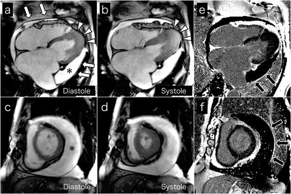
Fig. (3) Cine MR images on 4-chamber views (a and b) and short axis views (c and d) show a large amount of circumferential pericardial effusion (asterisks), thick visceral pericardium (white arrows), left ventricular apical myocardial hypertrophy with normal contraction (arrow-heads), and bilateral severe atrial dilatation, corresponding to echocardiography. Late gadolinium-enhanced images on 4-chamber views (e) and short axis views (f) show the visceral pericardium with high signal intensity (black arrows), indicating pericardial inflammation or fibrosis.