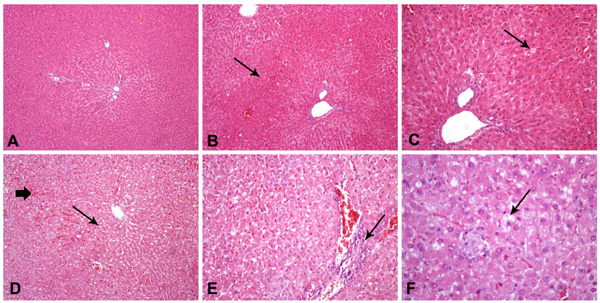
Fig. (1) Hematoxylin and eosin staining of hepatic tissue (200 and 400x).Control rats (Group A) with normal hepatocytes and liver architecture (A); Group HC(C) (HSF/HCH diet and vitamin E supplementation) rats with mild hepatic sinusoid congestion (B) and mild steatosis (C); Group HC(B) (HSF/HCH diet) rats with severe steatosis and hepatic sinusoid congestion (D), periportal (necro)-inflammatory infiltrates (E) and ballooning effect (F).