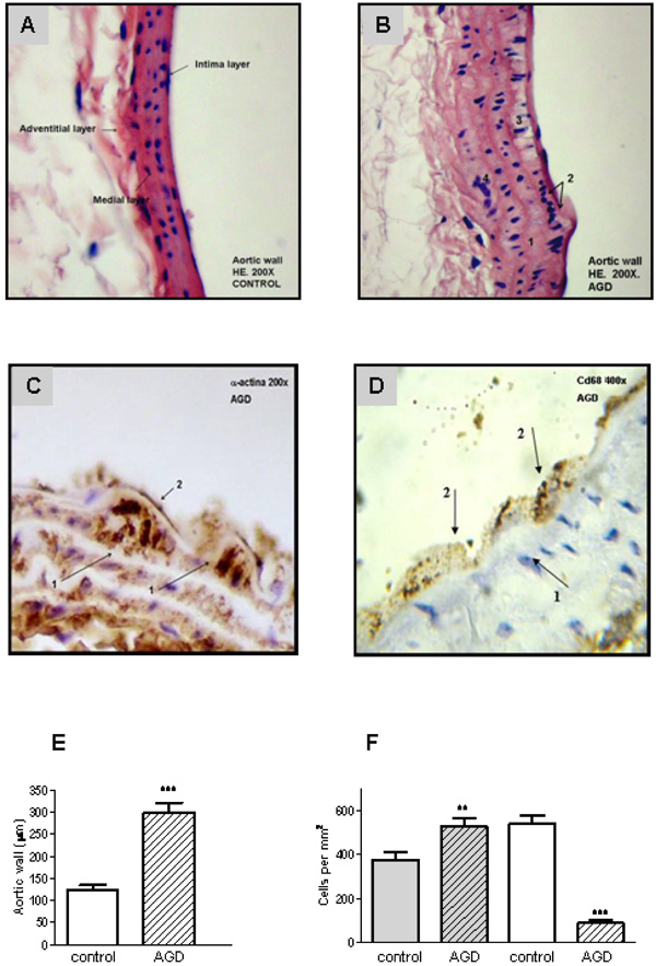
Fig. (2) Aortic lesions in AGD mice. Histological sections of aortic arteries, representative of n=10 sections from different animals of each
group, are shown. Panel A: Control aortic wall stained with Hematoxylin-eosin (HE) shows that all layers are conserved. Panel B: HE staining
of aortic wall from AGD mice: 1. rupture of the internal elastic layer; 2. migration of smooth muscle cells into the neo-intima; 3. vacuolization
of sub-intimal smooth muscle cells; 4. disruption of smooth muscle. Panel C: immunohistochemical staining with anti-a-actin antibody:
1-myoepithelial cells; 2-endothelial cells. Panel D: immunohistochemical staining anti-CD68 (macrophages). 1- myoepitelial cells, 2-
macrophage. Panel E: size of the intimal aortic wall. Panel F: average number of medial smooth muscle cells (SMCs). This value of intimal
cells was determined by counting nuclear profiles of two types of intimal cells: “synthetic”, undifferentiated cells (gray), and “contractile”,
differenciated cells: (white). Results are expressed as cells per unit area of intima (i.e., cells per square millimeter) in control and treated
(AGD) animals. Data represent mean ± SEM; n=3 in each group (Panel E and F), ** p< 0.01 and *** p<0.001