Fig. (1)
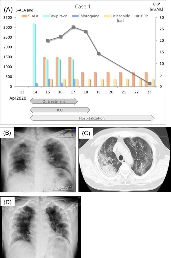
(A) Treatment course of Case 1. The ordinate on the left indicates the dose of 5-ALA, favipiravir, chloroquine, and ciclesonide (in µg) and that on the right indicates the concentration of CRP. The abscissa indicates the date. Administration of 1500 mg/day 5-ALA was initiated on April 15. The dose was reduced to 750 mg/day on April 18 because of the improvement of symptoms. CRP steeply decreased after April 18th. Periods of O2 treatment, ICU and hospitalization are indicated in the bottom. (B) Chest X-P of Case 1 taken at admission (April 14). Pneumonia in the right upper lobe and infiltrative shadow of the left hilum. (C) Chest CT scan taken at admission (April 14). Consolidation in the right lobe and ground-glass opacities in both lobes more localized at the periphery. (D) Chest X-P taken on April 23. Pulmonary lesions were improved, although there was a new shadow in the left lower lobe. Chest CT scan after recovery could not be obtained.