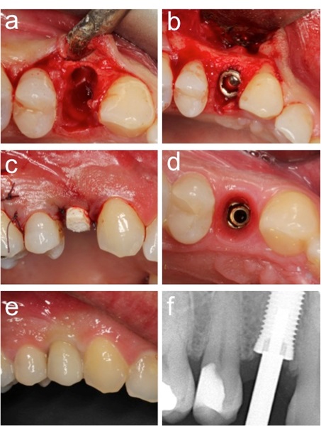
Fig. (2)
Clinical case. (a). Socket after extraction of premolar. (b). Tapered Neoss implant and bone substitute to fill voids around the implant collar. (c). Adjusted healing abutment. (d). Site after initial healing. (e). Intraoral radiograph at the one-year follow-up. (f). Final restoration..