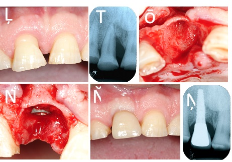
Fig. (3)
Clinical case. (a). Right central incisor with a deep pocket on the mesial aspect. (b). Preoperative radiograph. (c). Showing extraction socket. (d). Showing immediate implant placement in the extraction socket. The mesial defect has been packed with autogenous bone chips. (e). Intra-oral radiograph after one year of function. Note successful healing at the mesial aspect of the implants. (f). Final clinical appearance.