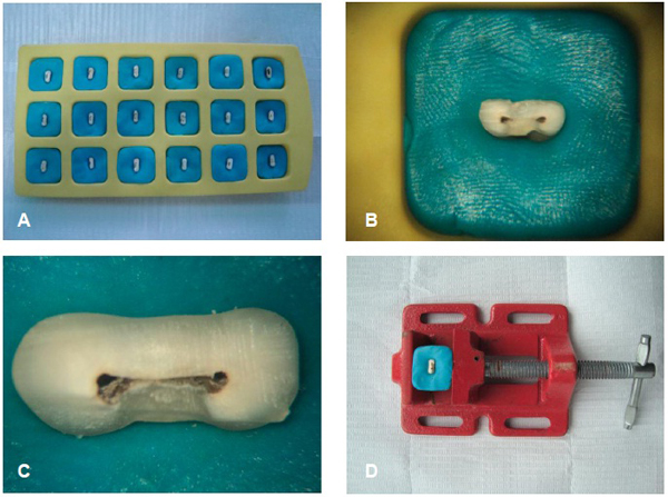
Fig. (2) (A). Specimens grouped into silicon blocks; (B). Magnification of the specimen with an increase of 8 X; (C). Magnification the specimen with 16 X; (D). Test specimens placed in a bench vise to instrumentation. For magnifying images was used microscope operative Alliance (São Carlos, São Paulo, Brazil).