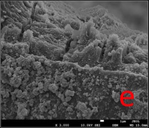
Fig. (4e)
SEM micrograph of a fractured site of dentin disc in DSC-AS Group: Crystalline layer on the surface and open tubule orifices at fractured site were visible (3,000X).