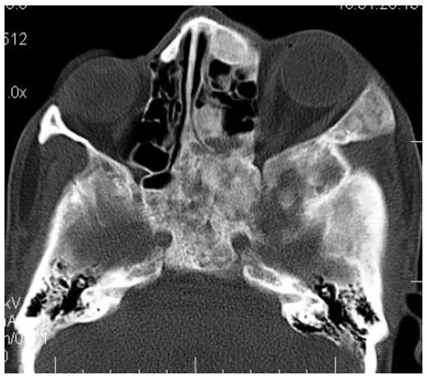
Fig. (1C)
Axial CT slice with bone window revealing involvement of temporal, zygomatic, frontal and ethmoid bones. Sphenoid bone was also involved with narrowing of the optic canal as well as orbital fissures.