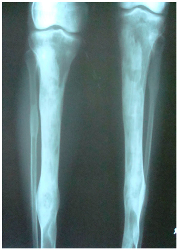
Fig. (2E)
Lateral AP view of Tibia with evidence of sclerosis of upper and mid shaft of tibia along with lytic areas in the sclerotic irregularity of the bone.