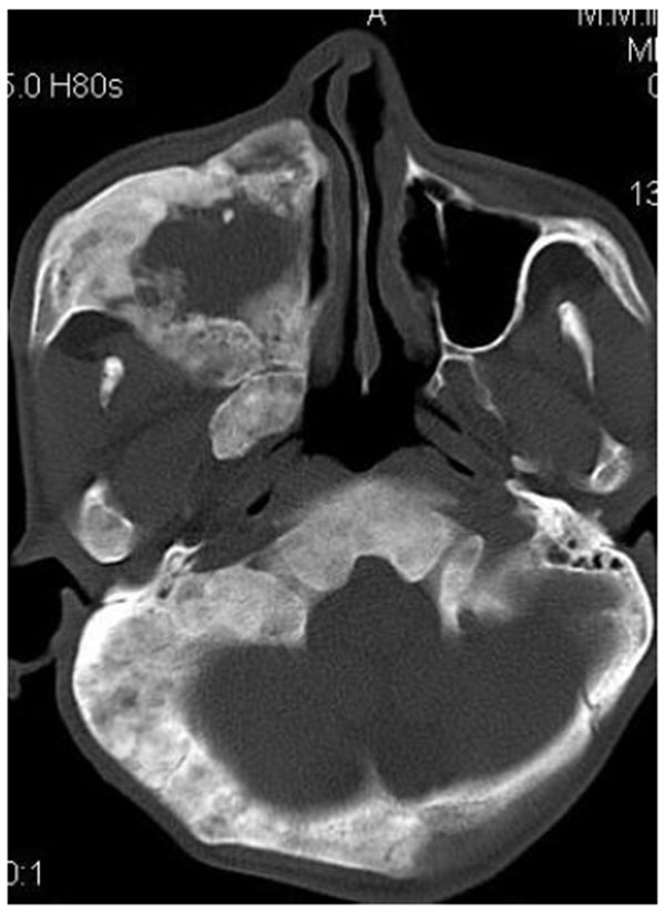
Fig. (2G)
Axial CT section showing the involvement of the maxillary sinus of right side. Further the zygomatic arch, zygoma, base of the cranium, mastoid process, pterygoid plates and sphenoid of right side were also involved. There was evidence of involvement of occipital condyles of both the sides and clivus.