Fig. (3D)
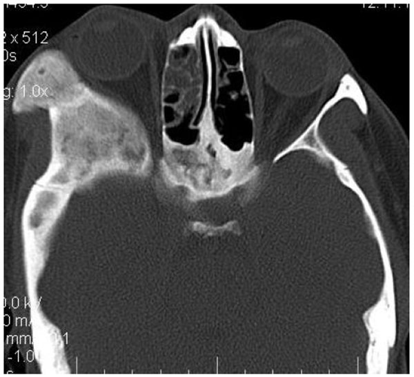
The Axial CT section revealing the involvement of the lateral wall of right orbit, right Zygomatic arch, zygoma, right pterygoid region with right craniaum. Further there was evidence of narrowing of the of the optic canal of right side.