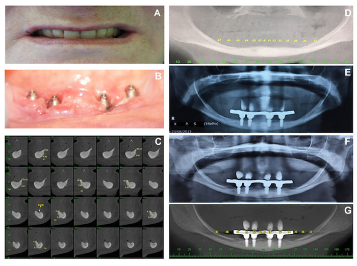
Fig. (1) A, Clinical image showing the loss of vertical dimension of occlusion used to establish the treatment plan; B: Implants placement after 1 week; C: Cone-beam computed tomography (CBCT) illustrating extremely reduced bone height and width; D: Panoramic vision of the reduced lower bone ridge; E: Panoramic examination showing the properly installed implants and bar without signs of peri-implant bone loss 30 days after surgery; F: Panoramic image 4 years after surgery; G: Panoramic image 56 months after surgery without signs of peri-implant bone loss or prosthesis maladaptation.