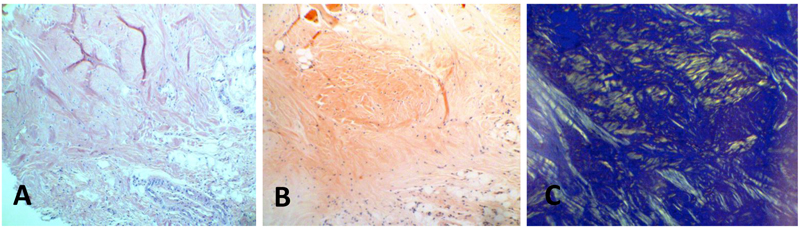
Fig. (1) (A) An incisional biopsy from the dorsal tongue of case number 9 was performed and histological examination showed an eosinophilic amorphous material in the connective tissue beneath the epithelium with a few inflammatory cells. (B) A red homogenous material with cracking artifact was visible in tissue sections stained with Congo red, and (C) The same area showed an apple green birefringence under polarized light. These findings were consistent with the diagnosis of amyloidosis.