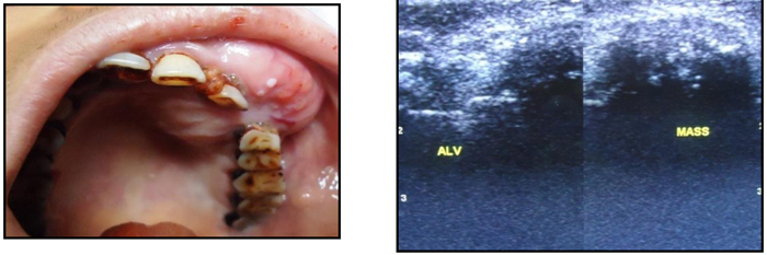
Fig. (2)
Benign tumor: Clinical swelling and Ultrasound showing a well defined hypoechoic maxillary mass with in between hyperechoic areas of calcifications.