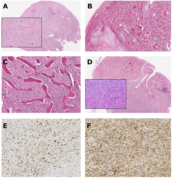
Fig. (4) Histological and immunohistochemical images. A. Histological image of fibrous component with moderate cellularity and atypical cells. B. Histological image showing enchondral ossification with cartilaginous tissue. C. Histological image of typical, well-differentiated irregular-woven boney trabeculae, focally arranged in parallel arrays in a background of spindle cell stroma (streamer pattern). D. Histological image of high-grade component with spindle cell sarcomatous fibrous tissue with mitotic nuclei. The sarcomatous tissue has grown infiltratively into the bone trabeculae. E and F. Immunohistochemical staining for MDM2 (E) and CDK4 (F) showing positive nuclear staining.