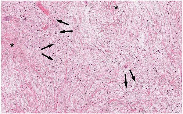
Fig. (3)
Peripheral odontogenic myxoma, patient 1. Histological examination showed randomly oriented spindled fibroblastic cells (black arrows) in a well-vascularized myxoid matrix (asterisks). H&E x 20.