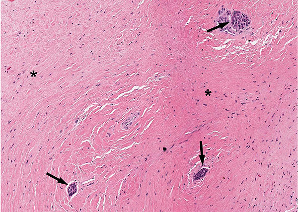
Fig. (4)
Peripheral odontogenic myxoma, patient 1. Histological examination showed scattered nests of inactive odontogenic epithelium (black arrows) surrounded by fibrous stroma (asterisks). H&E x 20.