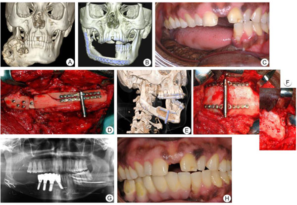
Fig. (2)
Case 2. Patient with ameloblastic carcinoma. A. CT-scan 3D image of the tumor mass at the right side of the mandible. B. CT-scan 3D image of the right side of the mandible reconstructed with fibula bone. C. Clinical photograph showing inadequate mandible height. D. Clinical photograph showing the placement of the distraction device. E-F. CT-scan 3D image (E) and clinical photograph (F) showing excellent ossification between the mandible and the distracted segment, six months after the consolidation period. G. Implants placement at the right side of the mandible. H. Clinical photograph showing implant-supported bridge at the right side of the mandible.