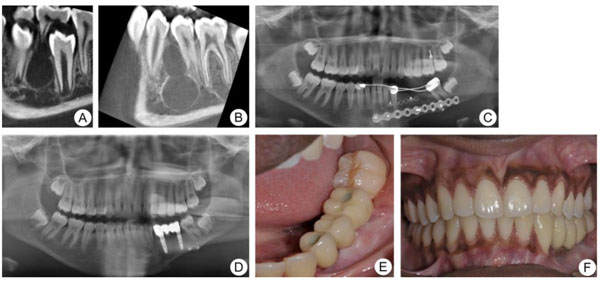
Fig. (3)
Case 3. Patient with multicystic ameloblastoma. A. CBCT image showing a cystic lesion between tooth 34 and 35. B. Six months postoperative control CBCT showing recurrence of multicystic lesion at the current location. C. Panoramic radiograph, three months after reconstruction with non-vascularized iliac bone graft at the left side of the mandible. D-F. One year postoperative panoramic radiograph (D) and clinical photographs (E & F) showing implant-supported fixed prosthesis at the left side of the mandible.