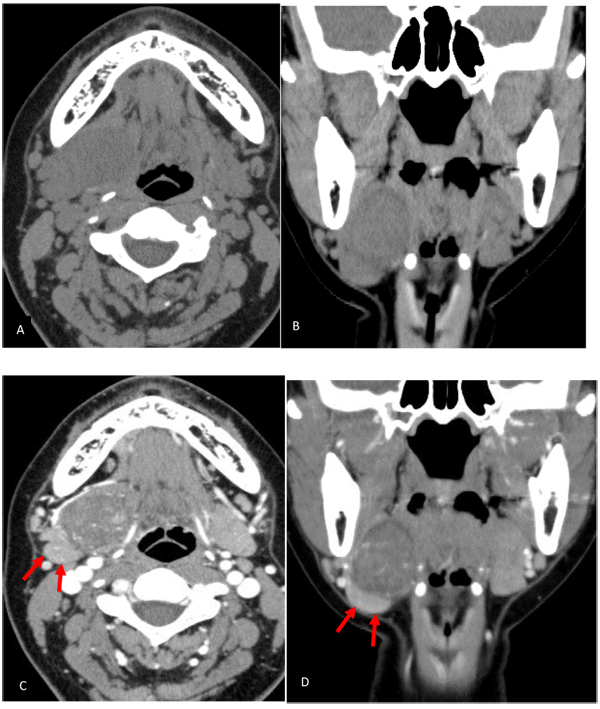
Fig. (1)
Non-contrast (A, B) and contrast-enhanced (C, D) CT images. The mass was located lateral to the genioglossus and hypoglossus muscles and the submandibular gland was compressed and inferiorly displaced (arrow). Contrast-enhanced CT demonstrated enhancement of intratumoral blood vessels.