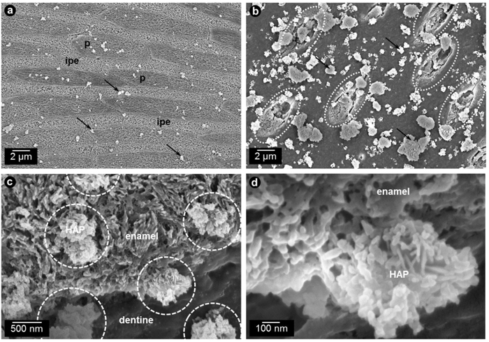
Fig. (3)
SEM-images of synthetic HAP-particles adhering to bovine enamel (a) and dentin (b) surfaces after having been applied in vitro. (a) Prismatic (p) and interprismatic enamel (ipe) can be clearly distinguished in this mildly etched sample (0.07 M phosphoric acid, 5 s), the synthetic particles are indicated by arrows. (b) Dentin surface of the same sample as (a), both exposed tubuli (encircled with dashed lines) and the bare surface are covered with synthetic HAP-particles (arrows). The affinity of the particles to the surface is higher for dentin than for enamel. (c) High magnification micrograph showing the structural similarity of HAP-particles (encircled with dashed lines) to the gently etched naturally formed HAP constituting the enamel. (d) Detail images show that the adhesion between enamel and particle is quite firm and individual crystallites at the interface can no longer be clearly associated to one or the other indicating a good mineral-mineral-interaction.