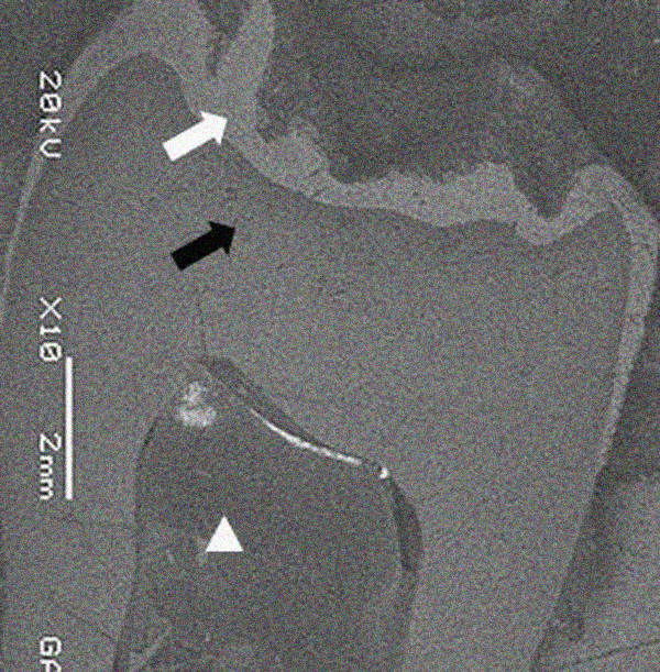
Fig. (5)
Scanning electron micrograph image of maxillary left third molar shows areas with irregular and thin enamel layer (white arrow), dentin (black arrow) and pulp chamber (white arrowhead).