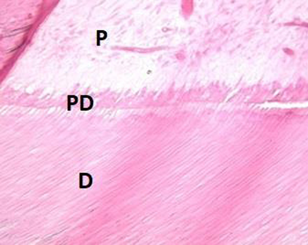
Fig. (6)
Histologic analysis of mandibular left third molar affected by AI revealed no defect and irregularity in tubular structure of dentin (Hematoxylin-eosin stain) (D=dentin, PD= primer dentin, P=pulp).