Fig. (1)
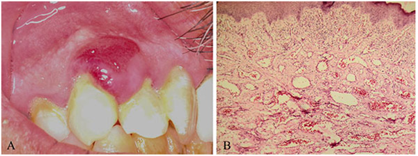
Morphological and histological views of Pyogenic granuloma. A) Clinical view of the pyogenic granuloma placed on upper attached gingiva between lateral and canine teeth. B) Section of the pyogenic granuloma excised out of one patient. Note the oral epithelium and the granulation tissue beneath (H&E staining, X200).