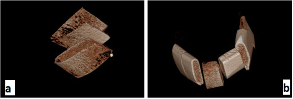
Fig. (1)
Three-dimensional reconstructed images of the second mandibular model taken by Cranex 3D ; a:in 4×6 cm
2
FOV and b: in 6×8 cm
2
FOV.