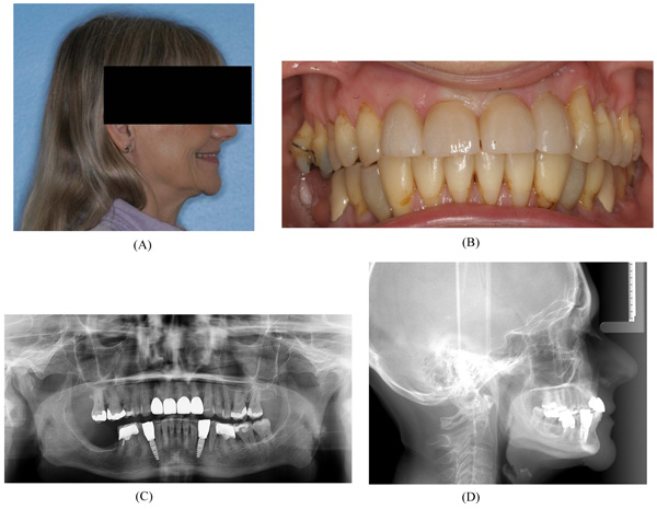
Fig. (8)
Final treatment result. (A) Clinical photo showing satisfying sagittal facial relations. (B) Intra-oral clinical photo showing the postoperative occlusion. (C) Orthopantomogram showing implant placement between the canines and first premolar. (D) Lateral cephalogram showing satisfying sagittal relations with normal incisor occlusion.