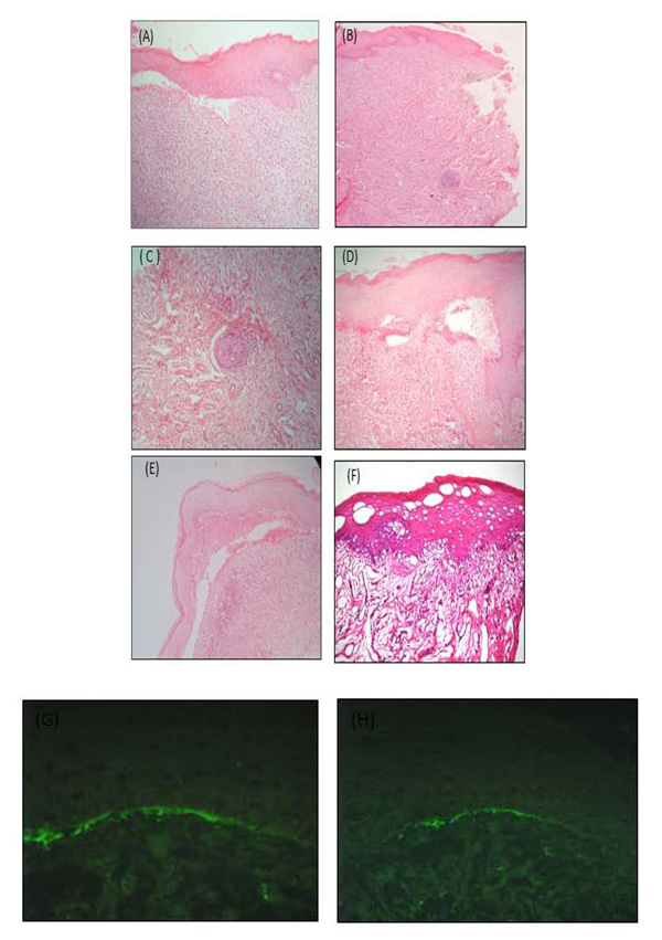
Fig. (2)
Histolopathological and direct immunofluorescence aspects of buccal mucosa and gingiva lesions. A) Histological cut of the buccal mucosa in HE staining (X100). B) Buccal mucosa fragment showing subepithelial lymphoplasmacytic inflammatory infiltrate and lymphoid follicle formation HEx100. C) Lymphoid follicle HEx40. D) Stratified keratinized squamous epithelium, chronically inflamed conjunctiva. E) Subepithelial cleft in the gingival region. F) Cornified hyperplasia and metaplasia of the stratified squamous epithelium, sparse apoptotic cells, pigmentary incontinence and moderate superficial interstitial lymphohistiocytic inflammatory infiltrate. (*) Artifacts: Retraction of both epithelial cells and collagen, leaving spaces. G) Focal granular deposit of IgM 2+ (on a scale of 1+ to 3+) in the basement membrane zone of the epidermis. H) The same in lower increase.