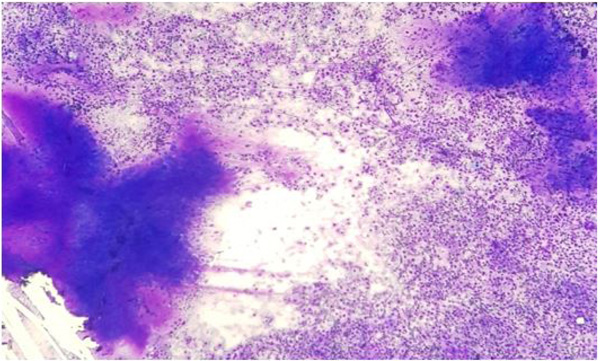
Fig. (1)
Pleomorphic adenoma – cytology: Smear showing singly scattered and irregular groups of epithelial cells with interspersed chondromyxoid stromal fragments and background population of myoepithelial cells (MGG, X100).