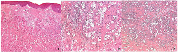
Fig. (3) A – Low power magnification showed nests and cords of odontogenic epithelial cells in the dense fibrous connective tissue under the mucosal epithelium (H&E, 50X original magnification). B. Varied size of epithelial islands composed by polyhedral cells with the presence of clear cells are also observed (H&E, 400X original magnification). C. Note the marked eosinophilic deposit interspersing the clear cell component (H&E, 400X original magnification).