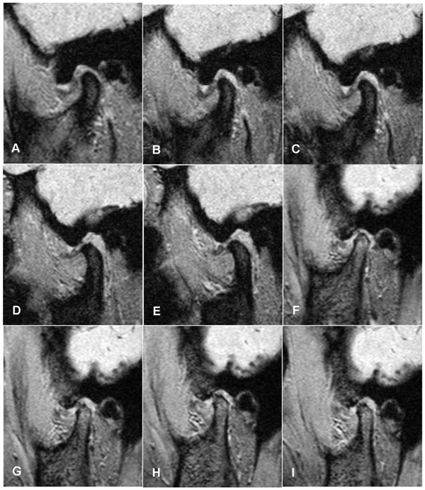
Fig. (5)
Simulation of the disc-condyle movement of TMJs with ADDWoR shown by Cine-MRI. FIESTA parameters, TR milliseconds/TE milliseconds=134.8.0/13.8 (A) In the chronic phase (over 6 months), at the closed-mouth position, the disc was significantly deformed. (B) At an open-mouth position of 1.0 cm, there was an onset of condylar rotation. The displaced disc was pushed forward during condylar rotation. (C) At an open-mouth position of 2.0 cm, condylar translation continued. (D) At an open-mouth position of 3.0 cm, there was condylar rotation, and the disc was pushed to the joint capsule and compressed. (E) At an open-mouth position of 4.0 cm, there was a rotational movement with a compressed disc, and the range of mandibular motion was usually not limited. (F) In the acute phase (within 6 months), at the closed-mouth position, the posterior band of the disc was anteriorly displaced relative to the condyle. (G) At an open-mouth position of 1.0 cm, there was an onset of condylar rotation, and the disc was pushed forward. (H) At an open-mouth position of 2.0 cm, condylar translation continued, and the disc was compressed and deformed. (I) At an open-mouth position of 3.0 cm, there was a condylar rotation, the disc was pushed to the joint capsule and was significantly compressed, and the range of mandibular motion was usually limited because of pain.