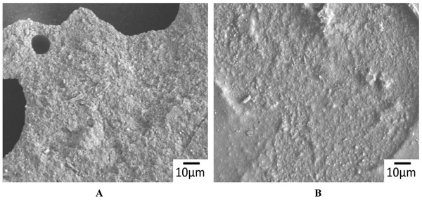
Figs. (5A & B)
SEM backscatter electron images of the surface of demineralized dentin after CAP I (plasma jet) Figs. (5A, B) and CAP II (DBD source) treatment Figs. (5A, B) and detachment (both 15 kV acceleration voltage, 500 x magnification). Similar to the non-demineralized samples vast areas of fractures within the adhesive/composite are visible after additional plasma irradiation.