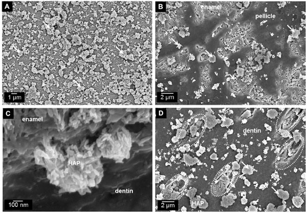
Fig. (2)
Scanning electron micrographs showing the attachment of synthetic HAP particles to the tooth surface in vitro (the hydroxyapatite phase of the particles was confirmed by X-ray powder diffraction). (A) Overview of attached synthetic HAP particles (arrows) as used in various oral care applications on clean bovine enamel substrate. (B) Synthetic HAP particles (arrows) attached to the surface of bovine teeth including enamel lesions and pellicle. (C) High magnification at the dentin-enamel junction shows a mineral–mineral interphase between HAP crystallites from synthetic particles and enamel. (D) Synthetic HAP particles attached to polished dentin surface including open tubules where particles (arrows) can be observed inside the open tubules. For more details see Fabritius-Vilpoux et al. [24].