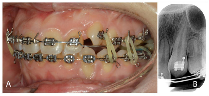
Fig. (1)
A) Pre-operative situation showing the ankylotic upper left canine, which was partially erupted and infraoccluded. B) Pre-operative radiograph showing reduced interradicular space between canine and first premolar.