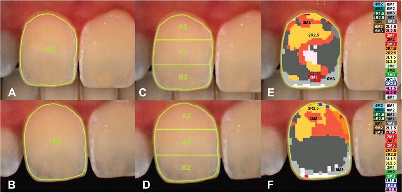
Fig. (3)
Example of case record clinical spectrophotometric documentation: (a-b) overall clinical
crown polarized image with closed and open occlusion; (c-d) evaluation of the three equal tooth area along the median axis: gingival, central, and incisal; with open and closed occlusion; (e-f) colour distribution and overall detailed mapping (Vita 3D Master scale) with closed and open occlusion.