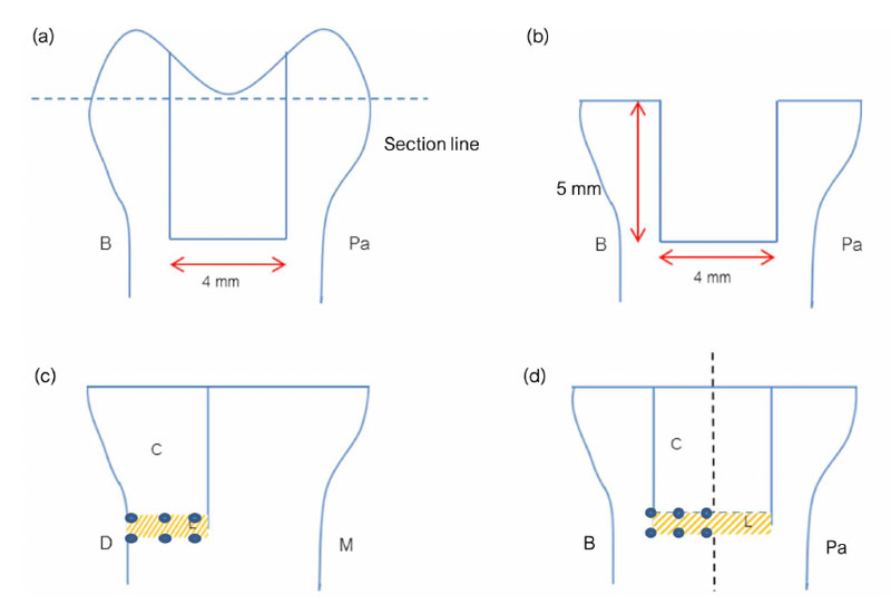
Fig. (1) The schematic drawing of sample preparation. The schematic drawing horizontally sectioned at the occlusal surface (a), cavity preparation after horizontally sectioned at the occlusal surface (b). Dots locate the gaps at liner-dentin interface and liner-resin composite interface of internal surface (c), and external surface (d). (C=Resin composite, L=Liner material, B=Buccal aspect, Pa= Palatal aspect, D=Distal aspect, M=Mesial aspect)