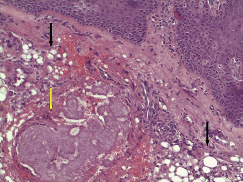
Fig. (4) H&E stain x 100: black arrows show silicon vacuoles within histiocytes in dermal tissue. In the middle (yellow arrow) some filamentous material is present, with the morphology of a cluster of Actinomyces colonies, forming “drusen”. Actinomyces colony filaments are colored by eosin stain in blue. The mycetoma is encircled with histiocytes of foreign body type and flattened fibrocytes.