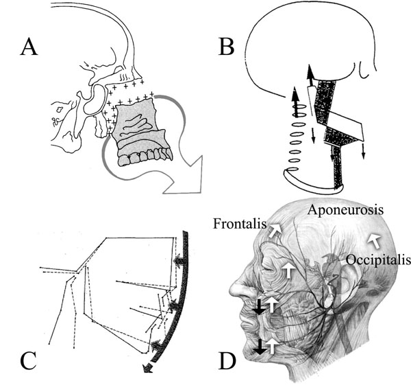
Fig. (1) (A) Displayed is the ventral and caudad growth direction of the ethmomaxillary complex Illustration from [1]. (B) Houston [16] combined his CFG model based on cervical vertebrae growth, with (C) the soft-tissue stretching of Solow [17] and cranial posture changes [114]. Illustrations from [16, 17]. Houston’s model cannot explain forward/counter-clockwise mandibular rotation. (D) An anatomical drawing of the aponeurotic tension model of craniofacial growth. Shown are: the force of gravity (black arrows) and CFMAS tension (white arrows); the frontalis muscle (frontalis), the occipitalis muscle (occipitalis) and the area between is the location of the galea aponeurotica (aponeurosis). The modiolus (muscular confluence joining the upper portion of the muscle mask with the lower portion) is found vertically between the black arrows overlying the cheek and chin. Illustration adapted from [115].