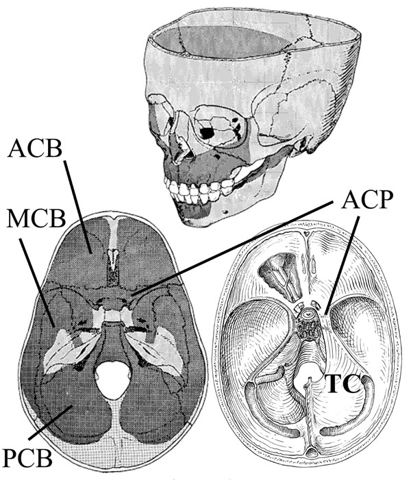
Fig. (4) Cranial resorption (dark stipple) and deposition patterns (light stipple) are illustrated on sectioned skulls. Areas of bone deposition are along the petrous portion of the temporal bone, crista galli/foramen cecum, between the occipital lobes, and sella turcica. The anterior (ACB), middle (MCB) and posterior (PCB) cranial fossa are shown (bottom left). The proposed normal distribution of facial bone resorption and deposition is shown (top). Bottom right displays the desmocranial lining of the cranial base; the lining is continuous with the falx cerebri but shown are the tentorium cerebelli (TC) and its attachment to the anterior clinoid process (ACP). This connection spans the SOS creating tension on the endocranial aspect of the SOS and contributing to differential growth of the SOS. Illustration from [48].