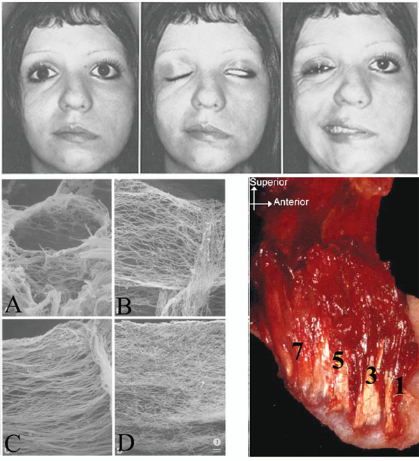
Fig. (5) (Top) Hemifacial paralysis is shown. Notice the tissue sag (left), and the resulting alar contour and relative vertical position between the paralyzed and normal sides (middle). There is arguably a more inferior position of the patient’s left orbit and eye. Photograph from [116] (Bottom left) There is a progressive age-related fiber thickening, densification and cross-linking of the connective tissue component of muscles (endomysium, perimysium, and epimysium) (bottom right) [62]. Age dependant directional restriction caused by the process requires further study. Photograph from [62]). (Bottom right) The original pterygomasseteric attachment may be preserved as the ramus grows posteriorly and new muscle attachment heads are developed; numbered from [1] initial/oldest attachment to [7] more recent attachment. Photograph from [103].