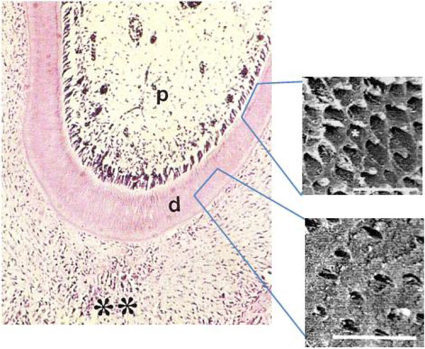
Fig. (1A) Photomicrograph of rat molars. Radicular region of GI molar showing the dental pulp (p), dentin (d) and periodontum (**). Odontoblastic palisade lining the dentin (Calibration bar: 50 µm). SEM reveals three-dimensional features including circumpulpal and peripheral dentin with dentinal tubules (*). (Calibration bar: 10 µm).