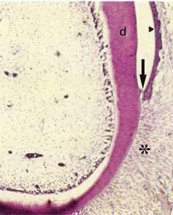
Fig. (1B) Photomicrograph of rat molars. In GII molars, no odontoblastic palisade or cell-free zone was observed. Note in the gingival sulcus, the sulcular epithelium (arrowhead) and loss of epithelial adhesion (arrow) within the junctional epithelium (HE, Calibration bar: 50 µm).