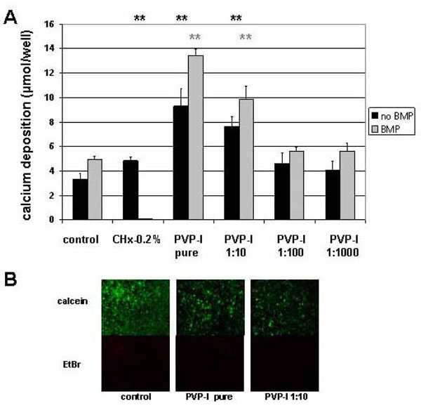
Fig. (2) Long-term effect of PVP-I treatment on preosteoblastic cells. Preosteoblastic cells were exposed to different concentrations of PVP-I on day 2 after seeding and evaluated 4 weeks later. (A) Alizarin-red staining method was used to determine mineral deposition. Dilutions of PVP-I of 1:100 and higher had no effect on the calcium deposition into the extracellular matrix. Exposure to undiluted or 1:10 diluted solutions of PVP-I, however, increased mineral deposition compared to the control untreated or respective BMP treated sample significantly (**) (**). (B) Live/dead staining revealed the presence of a majority of calcein stained living cells and a minority of EtBr stained dead cells in control samples. Although to a much lower extend living cells were also detected in samples treated with pure PVP-I or 1:10 dilutions. This minor fraction of surviving cells could account for the extended mineral deposition detected in the respective samples.