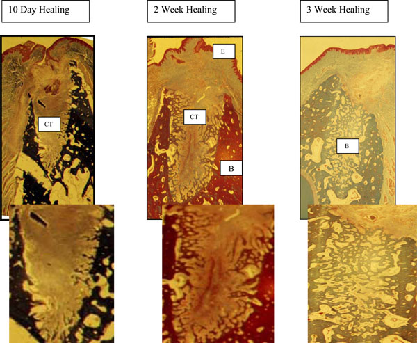
Fig. (1). PRFM alone and PRFM and Membrane 10 days - PRFM alone and PRFM and a membrane sockets were filled with an organized connective tissue (CT) and very little cellular infiltrate.2 weeks - complete epithelialization (E) of the wound had occurred. A well-organized fibrous connective tissue (CT) was seen with new bone growing from the entire periphery of the socket toward the center of the site.3 weeks - the sites were completely filled with new bone (B).