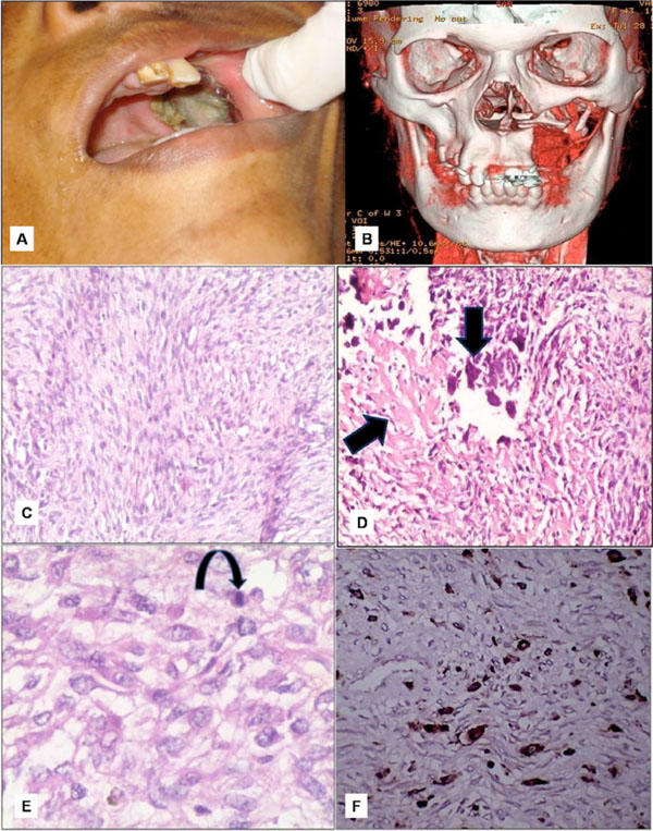
Fig. (5) A. Recurrent lesion measuring 2x3 cms in the maxillectomy region. B. 3D-imaging showing erosion of the floor of the left orbit. C. Storiform pattern of arrangement of fibroblasts with atypical; spindle cells. D. Osteoclasts like giant cells closely associated with tumour osteoid. E. Histiocytic type of cells admixed with atypical fibroblasts and atypical mitotic figures. F. Immunohistochemical CD68 marker study showing focal positivity for histiocytes.