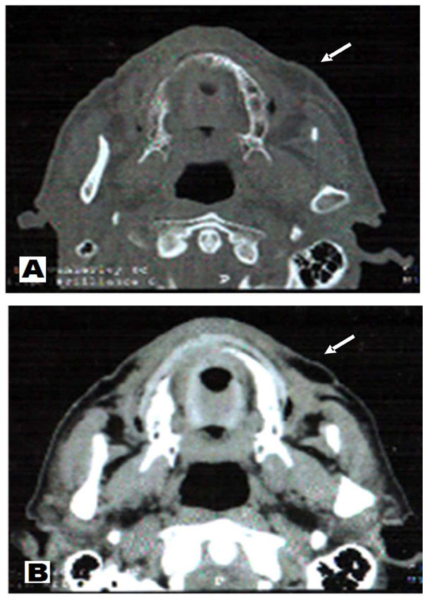
Fig. (2)
Axial CT scan showing a nodular image with sharp boundaries in part, measuring approximately 2.0x1.0 cm.
A
– CT soft tissue window.
B
– CT hard tissue window.